Atlas of Human Anatomy 7th Edition Pdf
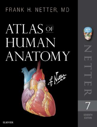
The only anatomy atlas illustrated by physicians, Atlas of Human Anatomy, 7th edition, brings you world-renowned, exquisitely clear views of the human body with a clinical perspective. In addition to the famous work of Dr. Frank Netter, you’ll also find nearly 100 paintings by Dr. Carlos A. G. Machado, one of today’s foremost medical illustrators. Together, these two uniquely talented physician-artists highlight the most clinically relevant views of the human body. In addition, more than 50 carefully selected radiologic images help bridge illustrated anatomy to living anatomy as seen in everyday practice.
Region-by-region coverage, including Muscle Table appendices at the end of each section.
Large, clear illustrations with comprehensive labels not only of major structures, but also of those with important relationships.
Tabular material in separate pages and additional supporting material as a part of the electronic companion so the printed page stays focused on the illustration.
Updates to the 7th Edition – based on requests from students and practitioners alike:
New Systems Overview section featuring brand-new, full-body views of surface anatomy, vessels, nerves, and lymphatics.
More than 25 new illustrations by Dr. Machado, including the clinically important fascial columns of the neck, deep veins of the leg, hip bursae, and vasculature of the prostate; and difficult-to-visualize areas like the infratemporal fossa.
New Clinical Tables at the end of each regional section that focus on structures with high clinical significance. These tables provide quick summaries, organized by body system, and indicate where to best view key structures in the illustrated plates.
More than 50 new radiologic images – some completely new views and others using newer imaging tools – have been included based on their ability to assist readers in grasping key elements of gross anatomy.
Updated terminology based on the international anatomic standard, Terminologia Anatomica, with common clinical eponyms included.
Student Consult access includes a pincode to unlock the complete enhanced eBook of the Atlas through Student Consult. Every plate in the Atlas?and over 100 Bonus Plates including illustrations from previous editions?are enhanced with an interactive label quiz option and supplemented with “Plate Pearls” that provide quick key points and supplemental tools for learning, reviewing, and assessing your knowledge of the major themes of each plate. Tools include 300 multiple choice questions, videos, 3D models, and links to related plates.
About the Author
Frank H. Netter was born in New York City in 1906. He studied art at the Art Students League and the National Academy of Design before entering medical school at New York University, where he received his Doctor of Medicine degree in 1931. During his student years, Dr. Netter’s notebook sketches attracted the attention of the medical faculty and other physicians, allowing him to augment his income by illustrating articles and textbooks. He continued illustrating as a sideline after establishing a surgical practice in 1933, but he ultimately opted to give up his practice in favor of a full-time commitment to art. After service in the United States Army during World War II, Dr. Netter began his long collaboration with the CIBA Pharmaceutical Company (now Novartis Pharmaceuticals). This 45-year partnership resulted in the production of the extraordinary collection of medical art so familiar to physicians and other medical professionals worldwide. Icon Learning Systems acquired the Netter Collection in July 2000 and continued to update Dr. Netter’s original paintings and to add newly commissioned paintings by artists trained in the style of Dr. Netter. In 2005, Elsevier Inc. purchased the Netter Collection and all publications from Icon Learning Systems. There are now over 50 publications featuring the art of Dr. Netter available through Elsevier Inc.
Dr. Netter’s works are among the finest examples of the use of illustration in the teaching of medical concepts. The 13-book Netter Collection of Medical Illustrations, which includes the greater part of the more than 20,000 paintings created by Dr. Netter, became and remains one of the most famous medical works ever published. The Netter Atlas of Human Anatomy, first published in 1989, presents the anatomic paintings from the Netter Collection. Now translated into 16 languages, it is the anatomy atlas of choice among medical and health professions students the world over.
The Netter illustrations are appreciated not only for their aesthetic qualities, but, more importantly, for their intellectual content. As Dr. Netter wrote in 1949 “clarification of a subject is the aim and goal of illustration. No matter how beautifully painted, how delicately and subtly rendered a subject may be, it is of little value as a medical illustration if it does not serve to make clear some medical point.? Dr. Netter’s planning, conception, point of view, and approach are what inform his paintings and what make them so intellectually valuable.
Frank H. Netter, MD, physician and artist, died in 1991.

